Brain phantoms are a creative solution for a challenging question: How do you tune an electromagnetic field to a patient without testing on the actual patient? Transcranial magnetic stimulation (TMS) is an application of electromagnetic research with the potential to change the way we treat migraines, depression, obsessive compulsive disorder and even conditions like schizophrenia and Parkinson’s disease.
Ravi Hadimani, Ph.D., associate professor of mechanical and nuclear engineering, leads a team of researchers who seek to use TMS to excite or inhibit brain neurons to alter specific brain functions and treat these conditions. This team includes faculty from VCU Health, including Mark Baron, M.D., professor of neurology and Kathryn Holloway, M.D., professor of neurosurgery, as well as outside collaborators like Joan Camprodon, M.D., associate professor of psychiatry at Harvard Medical School.
"The brain phantom is a first step,” says Hadimani, “Our ultimate goal is to 3D print a brain fabricated with biomaterial scaffolds and printed neurons that produce a stimulation response similar to neurons in our brain. This model would behave more realistically than current brain phantoms. Our future work involves collaborating with researchers who are able to print lab-grown neurons on biomaterial scaffolds or researchers who directly fabricate artificial neurons onto any scaffold."
Coils used in TMS are responsible for generating the electromagnetic field used in treatment. Individual coils are designed to treat specific diseases, but additional settings like current strength, number of pulses and coil direction are unique to each patient. Refining these settings on the actual patient is not feasible. Computer modeling is also inefficient because creating head models and running simulations from MRI scans of the brain’s complex structure are not spontaneous.
Hadimani and his team developed the brain phantom as a novel solution to this problem. In 2018, the first model was created by Hamzah Magsood, one of Hadimani’s Ph.D. students. The brain phantom is a physical model of a patient's brain designed to specifications obtained from MRI scans. Materials used in brain phantom construction are designed to replicate the electrical conductivity and electromagnetic permeability of different brain sectors. The result is a representation that, when connected to electrodes, provides instantaneous feedback to researchers calibrating TMS coils.
Elements of material science, electromagnetics and mechanical prototyping come together to create each brain phantom. The process starts with an MRI, which serves as a map for researchers designing the customized model. This is a careful process. Unlike other areas of the body with clear distinguishing features, like skin, muscle and bone, the brain has subtle differences between its many regions. Researchers must carefully distinguish between these areas to create an accurate brain phantom that will simulate a patient’s skin and skull as well as the brain’s gray and white matter.
A composite material of polymer and carbon nanotubes that exhibits electric properties similar to the human brain is the foundation for the brain phantom. Additive manufacturing, more commonly known as 3D printing, is used to create shells for different brain regions based on the patient’s MRI. This shell becomes a mold for the polymer and carbon nanotube solution. Once the brain phantom takes shape within the mold, it is placed within a solution that dissolves the casing, leaving only the brain phantom behind. The conductive parts of the brain phantom are dark because of the carbon nanotubes and non-conductive parts are lighter in color.
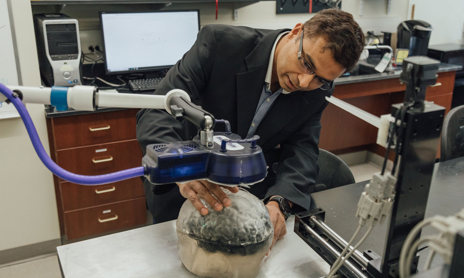
Electrodes are easily inserted into the brain phantom and provide feedback when an electromagnetic field from the TMS coil is applied. Adjustments to the strength, number of pulses of the field, and coil direction can then be made before applying the treatment to a patient.
Having recently received a patent for the brain phantom, Hadimani and Wesley Lohr, a senior biomedical engineering undergraduate, formed Realistic Anatomical Model (RAM) Phantom. The pair have been awarded both the Commonwealth Commercialization Fund Award and the Commonwealth Cyber Initiative Dreams to Reality Incubator Grant. RAM Phantom’s goal is to market brain phantom technology to the growing neuromodulation market, which also includes transcranial direct current stimulation and deep brain stimulation. The company will also aid in the development of advanced brain models that more accurately simulate the properties of the human brain.